One of the expectations embedded in the Singularity is the creation of a computer with the power of a human brain, and one necessary (it seems) task to that end is to reverse-engineer the human brain. That means researchers need to be able to examine it in minute detail, at extremely high resolutions. Which is what makes this development such an important step forward.
A multi-institutional consortium including Duke University has created startlingly crisp 3-D microscopic views of tiny mouse brains -- unveiled layer by layer -- by extending the capabilities of conventional magnetic resonance imaging.The important elements that I see in this report are the increased resolution of the MRI technology and the availability for study of the resulting images."These images can be more than 100,000 times higher resolution than a clinical MRI scan," said G. Allan Johnson, Duke's Charles E. Putman Distinguished Professor of radiology and professor of biomedical engineering and physics. He is first author of a report describing the innovations set for publication in the research journal NeuroImage
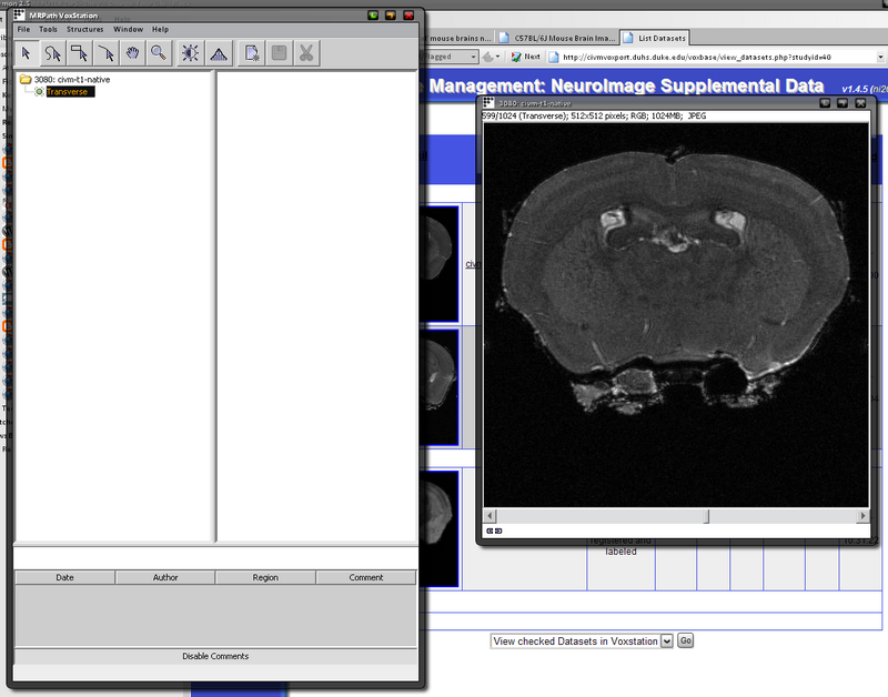
The consortium has developed the computer infrastructure to collect a rapidly growing library of 3-D mouse brain data, and make all the data available on the web http://tinyurl.com/3cgj6z. The goal is to use mouse brains as surrogates for human brains to study the connections between genes and brain structure. Investigators from all over the world are sending their models to Duke where the 3-D images are acquired in a standardized fashion and made available via high speed web connections.The specific research being done at Duke has to do with understanding the changes in phenotype (physical structure) that are associated with changes in genotype (gene expression). What happens if we don't allow this protein? How will that change the brain's structure? But this technology should also be useful to reverse-engineer the brain in order to simulate it effectively in a computer.High resolution magnetic resonance imaging -- which the researchers call "MRI histology" provides distortion-free 3-D images with superb ability to distinguish subtle tissue differences in the brain, according to Johnson.
"The specimen is still actually in the skull," he said. "It hasn't been cut by a knife. It has not been dehydrated and distorted as it would be in conventional histological techniques."
Using computer-guided statistical methods, the data can be segmented into more than 30 anatomical structures with quantitative volume measurements. These structures can then be computer-enhanced to produce color-coded and labeled volume renderings of selected anatomical details in 3-D, seen at any angle.
MRI scanning is also quicker and costs less than conventional histology, he said. MRI histology permits study of an entire brain, which would be prohibitively expensive using conventional methods.
The fact that you have access to these images via the Internet is a major step forward. Not that you or I can learn anything from them, but the right scientists can. That's important. The power of multiple minds at work!
[Source: Eureka Alert]
Singularity & The Price of Rice is updated daily; the easiest way to get your daily dose is by subscribing to our news feed. Stay on top of all our updates by subscribing now via RSS or Email.


0 comments :
Post a Comment